Animal Cell Microscope / Animal Cells And Plant Cells Cell Processes : Observe it under a compound microscope after staining and mounting.. One vital part of an animal cell is the nucleus. Air bubbles must be avoided in the sections. These regions of growth are good for studying the cell cycle because at any given time, you can find cells that are undergoing mitosis. It's the cell's brain, employing chromosomes to instruct other parts of the cell. Area where the chromatids of a chromosome are attached.
Observe it under a compound microscope after staining and mounting. Air bubbles must be avoided in the sections. Having observed the onion cell under the microscope, students will be able to learn the differences between animal and plant cells in addition to the function of the different parts of the cell. The mitochondria are the cell's powerplants, combining chemicals from our food with oxygen to create energy for the cell. In order to examine cells in the tip of an onion root, a thin slice of the root is placed onto a microscope slide and stained so the chromosomes will be visible.
Rana Ray Diagram Of Animal Cell Seen Through Electron Microscope Brainly In from hi-static.z-dn.net One vital part of an animal cell is the nucleus. Safranin is to be used to stain only the lignified tissues, over staining can be removed by washing in water. In order to examine cells in the tip of an onion root, a thin slice of the root is placed onto a microscope slide and stained so the chromosomes will be visible. Air bubbles must be avoided in the sections. Having observed the onion cell under the microscope, students will be able to learn the differences between animal and plant cells in addition to the function of the different parts of the cell. Observe it under a compound microscope after staining and mounting. Focus the slide under lower of microscope and then change to high power if needed precautions: Organize the spindle fibers to separate chromosomes during animal cell mitosis.
It helps in carrying out the functions such as respiration, nutrition, digestion, excretion etc.
Safranin is to be used to stain only the lignified tissues, over staining can be removed by washing in water. Focus the slide under lower of microscope and then change to high power if needed precautions: It helps in carrying out the functions such as respiration, nutrition, digestion, excretion etc. In order to examine cells in the tip of an onion root, a thin slice of the root is placed onto a microscope slide and stained so the chromosomes will be visible. One vital part of an animal cell is the nucleus. It's the cell's brain, employing chromosomes to instruct other parts of the cell. The mitochondria are the cell's powerplants, combining chemicals from our food with oxygen to create energy for the cell. Organize the spindle fibers to separate chromosomes during animal cell mitosis. So it is called as the structural and functional unit of life. Observe it under a compound microscope after staining and mounting. These regions of growth are good for studying the cell cycle because at any given time, you can find cells that are undergoing mitosis. Learn about onion root tip mitosis. The names of animal cell parts can be hard to.
The mitochondria are the cell's powerplants, combining chemicals from our food with oxygen to create energy for the cell. It's the cell's brain, employing chromosomes to instruct other parts of the cell. Safranin is to be used to stain only the lignified tissues, over staining can be removed by washing in water. The names of animal cell parts can be hard to. One vital part of an animal cell is the nucleus.
Anatomy And Physiology Of Animals The Cell Wikieducator from upload.wikimedia.org It's the cell's brain, employing chromosomes to instruct other parts of the cell. Organize the spindle fibers to separate chromosomes during animal cell mitosis. Air bubbles must be avoided in the sections. These regions of growth are good for studying the cell cycle because at any given time, you can find cells that are undergoing mitosis. The names of animal cell parts can be hard to. Area where the chromatids of a chromosome are attached. One vital part of an animal cell is the nucleus. Learn about onion root tip mitosis.
Learn about onion root tip mitosis.
Focus the slide under lower of microscope and then change to high power if needed precautions: Animal cells are packed with amazingly specialized structures. Observe it under a compound microscope after staining and mounting. Air bubbles must be avoided in the sections. Safranin is to be used to stain only the lignified tissues, over staining can be removed by washing in water. So it is called as the structural and functional unit of life. It helps in carrying out the functions such as respiration, nutrition, digestion, excretion etc. It's the cell's brain, employing chromosomes to instruct other parts of the cell. One vital part of an animal cell is the nucleus. Having observed the onion cell under the microscope, students will be able to learn the differences between animal and plant cells in addition to the function of the different parts of the cell. These regions of growth are good for studying the cell cycle because at any given time, you can find cells that are undergoing mitosis. Organize the spindle fibers to separate chromosomes during animal cell mitosis. In order to examine cells in the tip of an onion root, a thin slice of the root is placed onto a microscope slide and stained so the chromosomes will be visible.
Learn about onion root tip mitosis. Area where the chromatids of a chromosome are attached. One vital part of an animal cell is the nucleus. Having observed the onion cell under the microscope, students will be able to learn the differences between animal and plant cells in addition to the function of the different parts of the cell. These regions of growth are good for studying the cell cycle because at any given time, you can find cells that are undergoing mitosis.
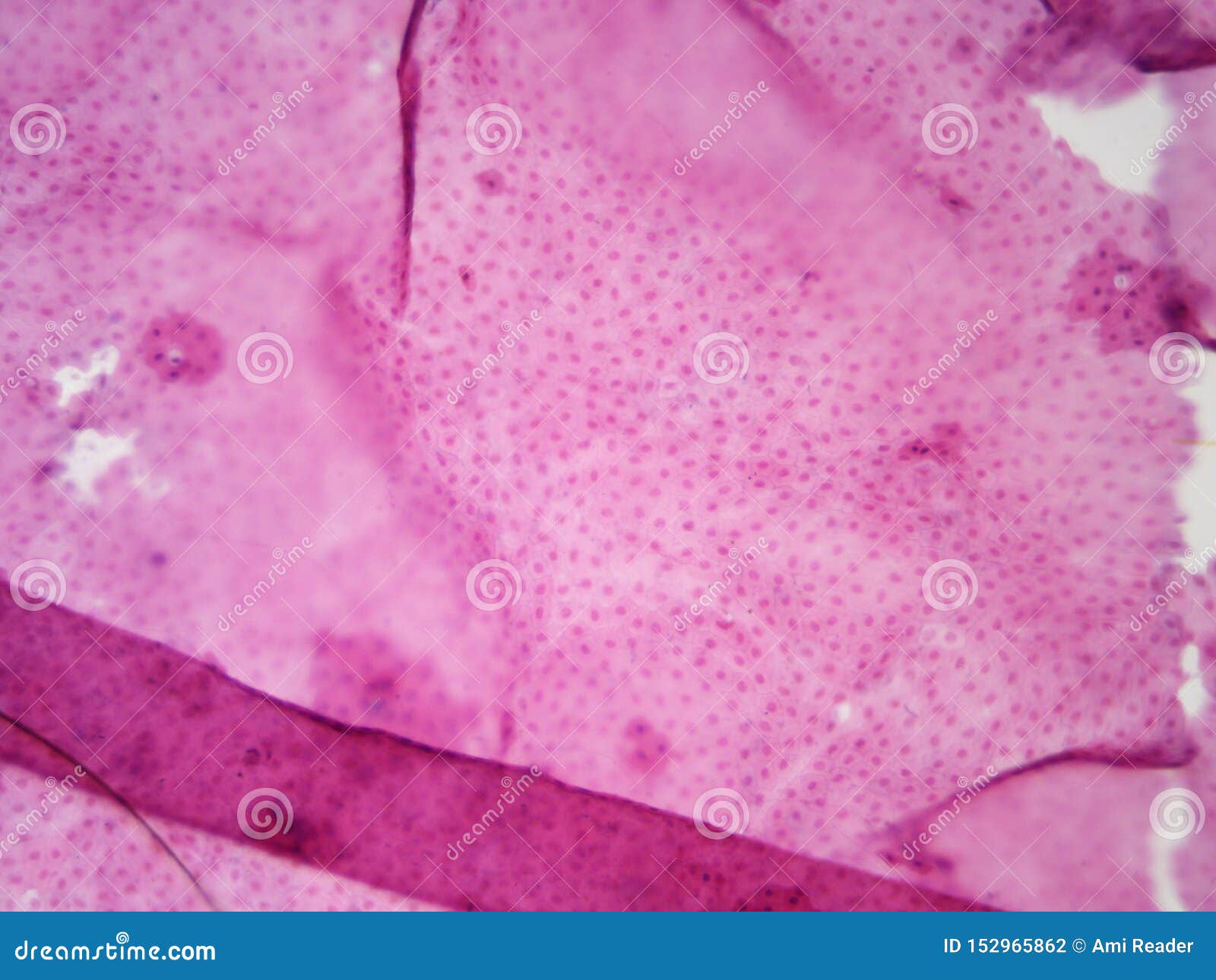
Typical Animal Cell Center 100x Stock Photo Image Of 100x School 152965862 from thumbs.dreamstime.com So it is called as the structural and functional unit of life. Learn about onion root tip mitosis. Observe it under a compound microscope after staining and mounting. These regions of growth are good for studying the cell cycle because at any given time, you can find cells that are undergoing mitosis. The names of animal cell parts can be hard to. In order to examine cells in the tip of an onion root, a thin slice of the root is placed onto a microscope slide and stained so the chromosomes will be visible. Organize the spindle fibers to separate chromosomes during animal cell mitosis. The mitochondria are the cell's powerplants, combining chemicals from our food with oxygen to create energy for the cell.
So it is called as the structural and functional unit of life.
It's the cell's brain, employing chromosomes to instruct other parts of the cell. Learn about onion root tip mitosis. In order to examine cells in the tip of an onion root, a thin slice of the root is placed onto a microscope slide and stained so the chromosomes will be visible. These regions of growth are good for studying the cell cycle because at any given time, you can find cells that are undergoing mitosis. So it is called as the structural and functional unit of life. Air bubbles must be avoided in the sections. Having observed the onion cell under the microscope, students will be able to learn the differences between animal and plant cells in addition to the function of the different parts of the cell. Area where the chromatids of a chromosome are attached. Animal cells are packed with amazingly specialized structures. Organize the spindle fibers to separate chromosomes during animal cell mitosis. Focus the slide under lower of microscope and then change to high power if needed precautions: Observe it under a compound microscope after staining and mounting. It helps in carrying out the functions such as respiration, nutrition, digestion, excretion etc.


0 Comments